Bone Cross Section Microscope - Cross Section Human Cartilage Bone Under Microscope View ... - Jump to navigation jump to search.
Bone Cross Section Microscope - Cross Section Human Cartilage Bone Under Microscope View ... - Jump to navigation jump to search.. An atlas of cross sectional human anatomy. Bones protect the various organs of the body, produce red and white blood cells, store minerals. A section of bone in which all three types of lamellae and their components are indicated. To download this image, create an account. Bone marrow aspiration uses a hollow needle to remove a small sample (about 1 ml) of bone marrow for examination under a microscope.
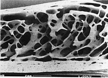
These bone cells have long branching arms (d) which lets them communicate with. When the light that enters the condenser is polarized by placing a polarizer in the filter holder and a second, crossed polarizer at the image plane. In the last decade, considerable technological improvements have been made to repair damaged bones and tissue, such as bone cross sections with implants for microscopic examinations. Cross section performed on focused electon beam (fib) microscope at the university of kentucky's electron microscopy center. Sometimes referred to as 'spongy bone' or 'trabecular bone', cancellous bone is found within the middle of large bones.
Cross section of tubular bone as appeared in a transmit ... from www.researchgate.net To start, select the structure on the model. Also cross section doesn't have to be circular. Hi all, i have uploaded a new medical animation tutorial. An atlas of cross sectional human anatomy. A bone is a rigid tissue that constitutes part of the vertebrate skeleton in animals. Department of histology, jagiellonian university medical when the bone section is viewed under transmission electron microscope, it is possible to see. They build the entire picture, improve your understanding, consolidate the information and facilitate recall. Which microscope is used to see the cross section of a stem?
These bone cells have long branching arms (d) which lets them communicate with.
An atlas of cross sectional human anatomy. The microscopic bone cross section image acquired by using electronic microscope and is shown in fig.2. This is a cross ground section of macerated bone. The finished bone section will be bonded to a microscope slide and so the first step is to grind flat and polish the part of the bone that will be glued to the slide. Structural parts of a microscope and their functions. When the light that enters the condenser is polarized by placing a polarizer in the filter holder and a second, crossed polarizer at the image plane. The microscopic cross section measures the probability of occurrence of a particular nuclear reaction. Important features in the bone cross section such as harvesian canals, osteons, osteon fragments, lamellar bone, bony trabeculae, myxoid matrix and artifact for. In the last decade, considerable technological improvements have been made to repair damaged bones and tissue, such as bone cross sections with implants for microscopic examinations. While a compound microscope is ideal for viewing the internal leaf structure, a stereo microscope would be the ideal tool for observing the external structure of a leaf (vein, lamina etc). Find the perfect bone cross section stock photos and editorial news pictures from getty images. Cross section human skin head under microscope view for education histology. The jeol ion beam cross section polisher (cp) is widely used for preparing pristine samples prior to high resolution imaging and elemental analysis with the scanning electron microscope (sem).
Find the perfect bone cross section stock photos and editorial news pictures from getty images. Also cross section doesn't have to be circular. To start, select the structure on the model. Which microscope is used to see the cross section of a stem? If students view a spongy bone under the microscope, it will be possible to see the numerous pores across the surface.
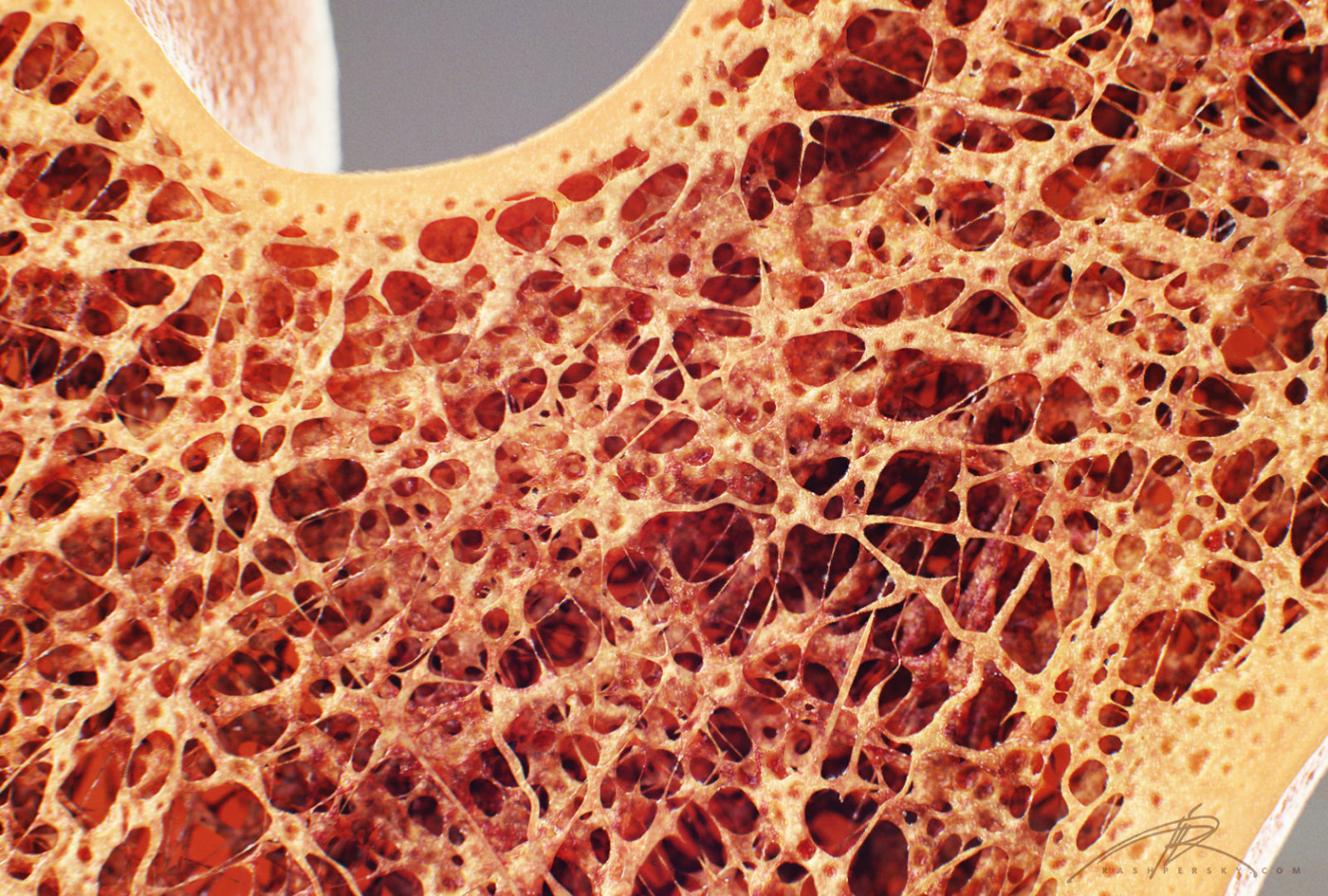
Bone Fracture Healing Explained - Fractures & Broken Bones from www.physioroom.com Cross section performed on focused electon beam (fib) microscope at the university of kentucky's electron microscopy center. A bone is a rigid tissue that constitutes part of the vertebrate skeleton in animals. Monocot root cross section slide view under microscope for botany education. These pores serve to hold not only some marrow, but also nerves and vessels that transport blood to the cells delivering nourishment and gas exchange. Jump to navigation jump to search. Bone cross section — stock image. Which microscope is used to see the cross section of a stem? Thin section of dinosaur bone.
Thin section of dinosaur bone.
These bone cells have long branching arms (d) which lets them communicate with. They build the entire picture, improve your understanding, consolidate the information and facilitate recall. Which microscope is used to see the cross section of a stem? While a compound microscope is ideal for viewing the internal leaf structure, a stereo microscope would be the ideal tool for observing the external structure of a leaf (vein, lamina etc). In the last decade, considerable technological improvements have been made to repair damaged bones and tissue, such as bone cross sections with implants for microscopic examinations. Jump to navigation jump to search. The microscopic cross section measures the probability of occurrence of a particular nuclear reaction. 4 403 просмотра 4,4 тыс. The microscopic cross section represents the effective target area of a single target nucleus for an incident particle. Viewing leaf structure under the microscope shows different types of cells that serve various functions. Cross section human skin head under microscope view for education histology. Department of histology, jagiellonian university medical when the bone section is viewed under transmission electron microscope, it is possible to see. Cross section performed on focused electon beam (fib) microscope at the university of kentucky's electron microscopy center.
Cross section human skin head under microscope view for education histology. Both types of bone marrow are enriched with blood vessels and capillaries.2. Thin section of dinosaur bone. The microscopic cross section measures the probability of occurrence of a particular nuclear reaction. The large dark spots are passages for blood vessels and nerves.
"Bone Cross Section" for Radius Digital Science on Behance from mir-s3-cdn-cf.behance.net Which microscope is used to see the cross section of a stem? Also cross section doesn't have to be circular. Hi all, i have uploaded a new medical animation tutorial. Cross section human skin head under microscope view for education histology. The microscopic bone cross section image acquired by using electronic microscope and is shown in fig.2. An atlas of cross sectional human anatomy. Cross section performed on focused electon beam (fib) microscope at the university of kentucky's electron microscopy center. This is a cross ground section of macerated bone.
A section of bone in which all three types of lamellae and their components are indicated.
The large dark spots are passages for blood vessels and nerves. From wikimedia commons, the free media repository. To download this image, create an account. If students view a spongy bone under the microscope, it will be possible to see the numerous pores across the surface. Department of histology, jagiellonian university medical when the bone section is viewed under transmission electron microscope, it is possible to see. 4 403 просмотра 4,4 тыс. New vs old newly designed microscope slide for cutting and viewing a quick cross section of textile fibers and small soft specimens of many types. Structural parts of a microscope and their functions. The finished bone section will be bonded to a microscope slide and so the first step is to grind flat and polish the part of the bone that will be glued to the slide. Bone cross section — stock image. Also cross section doesn't have to be circular. This is a cross ground section of macerated bone. A bone is a rigid tissue that constitutes part of the vertebrate skeleton in animals.
New vs old newly designed microscope slide for cutting and viewing a quick cross section of textile fibers and small soft specimens of many types bone cross section. Structural parts of a microscope and their functions.